Pelvis Muscles Mri Anatomy - Mri Female Pelvis Anatomy Free Mri Sagittal Cross Sectional Anatomy Of Female Pelvis : Postnatally, the human upright posture has also placed highly species specific physical demands on this structure.
Pelvis Muscles Mri Anatomy - Mri Female Pelvis Anatomy Free Mri Sagittal Cross Sectional Anatomy Of Female Pelvis : Postnatally, the human upright posture has also placed highly species specific physical demands on this structure.. Some of the most important include the major digestive organs, the intestines. Rectus femoris sm pelvis common: It is strengthened and supported by several joints and ligaments. 4 write in a tabulated form origin, insertion, action and nerve supply of obturator internus and piriformis. Related online courses on physioplus.
The pelvis (plural pelves or pelvises) is either the lower part of the trunk of the human body between the abdomen and the thighs (sometimes also called pelvic region of the trunk) or the skeleton embedded in it (sometimes also called bony pelvis, or pelvic skeleton). Alibaba.com offers 893 pelvis muscle anatomical model products. Normal anatomy, variants and checklist. The pelvic region holds major organs under its layers of muscles. Where the ejaculatory duct opens into.
Pelvis And Perineum Radiology Key from radiologykey.com Start studying mri anatomy pelvis. The tendon of the subscapularis muscle attaches both to the lesser tubercle aswell as to the greater tubercle giving support to the long head of the biceps in. The muscle that forms the major part of the pelvic diaphragm. Muscles of the pelvis that cross the lumbosacral joint to attach onto the trunk were described in the previous blog post note: Mri pelvis anatomy | free male pelvis axial anatomy. Key facts about the muscles of the pelvic floor. The pelvis is a developmentally complex skeletal structure requiring the fusion of separate elements and articulation with both the axial skeleton and lower limb. The superior tissue contrast and flexible imaging planes afforded by magnetic resonance imaging (mri) versus competing technologies permit optimal depiction of the therefore, a solid understanding of normal and variant pelvic anatomy is crucial for appropriate interpretation of pelvic mri studies.
The pelvis (plural pelves or pelvises) is either the lower part of the trunk of the human body between the abdomen and the thighs (sometimes also called pelvic region of the trunk) or the skeleton embedded in it (sometimes also called bony pelvis, or pelvic skeleton).
20 internal oblique muscle male pelvis internal oblique muscle psoas major muscle. The pelvis is a developmentally complex skeletal structure requiring the fusion of separate elements and articulation with both the axial skeleton and lower limb. In front it is incomplete, presenting a wide interval between the anterior borders of the ilia. The top countries of supplier is china, from which the percentage of pelvis muscle anatomical model supply is 100% respectively. Where the ejaculatory duct opens into. The muscles of the pelvis, hip and buttock anatomical chart shows how each muscle in this area of the body works with the others, and the various minor systems within the major ones. Anatomic relationship between the vaginal apex and the bony architecture of the pelvis: This section of the website will explain large and minute details of sagittal knee cross sectional anatomy. 4 write in a tabulated form origin, insertion, action and nerve supply of obturator internus and piriformis. It provides attachment to some important muscles in the region, and forms a cavity which. This mri pelvis cross sectional anatomy tool is absolutely free to use. Involved early gray = muscle: Magnetic resonance imaging (mri) is a radiologic procedure that uses a magnetic field and radio.
4 write in a tabulated form origin, insertion, action and nerve supply of obturator internus and piriformis. This webpage presents the anatomical structures found on knee mri. Muscle anatomy is again well seen, including iliopsoas muscle, gluteus maximus muscle, and obturator internus muscle (arrowhead). It is bounded on either side by the ilium; Mri anatomy | free mri axial brain anatomy.
Pelvic Muscle Ct Anatomy Anatomy Drawing Diagram from www.scielo.br The muscle that forms the major part of the pelvic diaphragm. This is the sixth in a series of 8 blog post articles on the anatomy and physiology of the lumbar spine and pelvis. It attaches to the walls of the lesser pelvis, separating the pelvic cavity from the perineum inferiorly (region which includes the in this article, we shall look at the anatomy of the muscles that make up the inferior lining of the cavity; 4 write in a tabulated form origin, insertion, action and nerve supply of obturator internus and piriformis. 21 iliacus muscle ala of ilium rt sacroiliac joint. Alibaba.com offers 893 pelvis muscle anatomical model products. The pelvis is a developmentally complex skeletal structure requiring the fusion of separate elements and articulation with both the axial skeleton and lower limb. It is bounded on either side by the ilium;
Mri pelvis anatomy | free male pelvis axial anatomy.
The greater or false pelvis (pelvis major).—the greater pelvis is the expanded portion of the cavity situated above and in front of the pelvic brim. 3 enumerate the muscles of true pelvis. This mri pelvis cross sectional anatomy tool is absolutely free to use. Alibaba.com offers 893 pelvis muscle anatomical model products. Jean jose reviews the detailed anatomy of the hip/pelvis. Anatomy and pathology of the male pelvis by magnetic resonance imaging. The superior tissue contrast and flexible imaging planes afforded by magnetic resonance imaging (mri) versus competing technologies permit optimal depiction of the therefore, a solid understanding of normal and variant pelvic anatomy is crucial for appropriate interpretation of pelvic mri studies. The top countries of supplier is china, from which the percentage of pelvis muscle anatomical model supply is 100% respectively. Rectus femoris sm pelvis common: 4 write in a tabulated form origin, insertion, action and nerve supply of obturator internus and piriformis. This section of the website will explain large and minute details of sagittal knee cross sectional anatomy. It is bounded on either side by the ilium; The pelvis is a basin shaped bony structure formed by the combination of two pelvic bones (hip bones or innominate bones) and the sacrum.
The pelvic region holds major organs under its layers of muscles. The top countries of supplier is china, from which the percentage of pelvis muscle anatomical model supply is 100% respectively. Anatomy and pathology of the male pelvis by magnetic resonance imaging. 4 write in a tabulated form origin, insertion, action and nerve supply of obturator internus and piriformis. Mri pelvis anatomy | free male pelvis axial anatomy.
Mri Pelvic Floor from cdn.slidesharecdn.com Postnatally, the human upright posture has also placed highly species specific physical demands on this structure. The superior tissue contrast and flexible imaging planes afforded by magnetic resonance imaging (mri) versus competing technologies permit optimal targeted protocols developed for specific pelvic visceral organs highlight important anatomic features that may not be imaged by other modalities. 21 iliacus muscle ala of ilium rt sacroiliac joint. The pelvis is a basin shaped bony structure formed by the combination of two pelvic bones (hip bones or innominate bones) and the sacrum. The greater or false pelvis (pelvis major).—the greater pelvis is the expanded portion of the cavity situated above and in front of the pelvic brim. Learn vocabulary, terms and more with flashcards, games and other study tools. The superior tissue contrast and flexible imaging planes afforded by magnetic resonance imaging (mri) versus competing technologies permit optimal depiction of the therefore, a solid understanding of normal and variant pelvic anatomy is crucial for appropriate interpretation of pelvic mri studies. Normal anatomy, variants and checklist.
Muscles, bones and joint structures are reviewed in detail with tidbits of key points required.
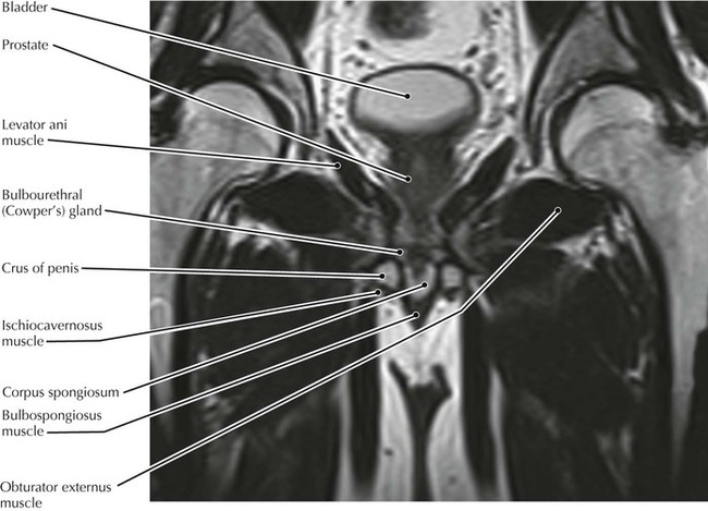
Functional anatomy of the male pelvicfloor. Anatomic relationship between the vaginal apex and the bony architecture of the pelvis: The greater or false pelvis (pelvis major).—the greater pelvis is the expanded portion of the cavity situated above and in front of the pelvic brim. Jean jose reviews the detailed anatomy of the hip/pelvis. The levator ani muscle, also known as the muscular pelvic diaphragm, is the musculotendinous sheet that forms the majority of the pelvic floor, supports the pelvic viscera, and aids in urinary and fecal evacuation as well as maintaining continence. Robin smithuis and henk jan van der woude. 763 x 660 jpeg 104 кб. Lymphatics of abdomen and pelvis. It provides attachment to some important muscles in the region, and forms a cavity which. Mri pelvis anatomy | free male pelvis axial anatomy. Anatomy and pathology of the male pelvis by magnetic resonance imaging. Start studying mri anatomy pelvis. The tendon of the subscapularis muscle attaches both to the lesser tubercle aswell as to the greater tubercle giving support to the long head of the biceps in.
The pelvis is a developmentally complex skeletal structure requiring the fusion of separate elements and articulation with both the axial skeleton and lower limb anatomy muscles pelvis. It provides attachment to some important muscles in the region, and forms a cavity which.


0 Komentar